TRABAJOS de CASUÍSTICA y
CASOS CLÍNICOS
Linfangitis carcinomatosa pulmonar: presentación como
neumopatía aguda. reporte de dos casos.
Pulmonary
carcinomatous lymphangitis: presentation as acute
pneumopathy.report of two cases.
Ré DP*, Cazaux A, Cambursano VH,
Zaya A, Cortés JR.
Revista Facultad de Ciencias
Medicas 2013; 70(1):31-33
Hospital Rawson. Bajada Pucará
2025, Córdoba, Argentina. Tel: 0351-6805976
*E-mail de contacto:
danila_re@hotmail.com
Resumen
Introducción: La linfangitis carcinomatosa(LC)
representa el 6-8% de las metástasis pulmonares. Existe
evidencia de que puede ser una condición oncológicamente
tratable, con impacto sobre la progresión de la disnea y
lesiones radiológicas, con mejoría de la sobrevida. Se
describen dos casos con el objetivo de inducir la sospecha
de esta entidad ante un paciente con datos de neumopatía
aguda. Métodos: Caso 1: mujer 32 años, disnea progresiva y
tos de dos semanas de evolución sin respuesta a
antibióticos. Taquipnea, aumento del trabajo respiratorio,
crepitantes bibasales. Insuficiencia respiratoria.
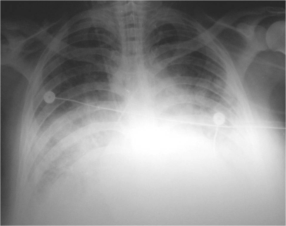
Radiografía: radiopacidadalvéolointersticialbibasal. Se
inicia tratamiento para neumonía grave de la comunidad.
Evoluciona desfavorablemente con requerimiento de ARM y
desenlace fatal. Caso 2: mujer 46 años, disnea progresiva y
tos de una semana de evolución. Taquipnea, subcrepitantes
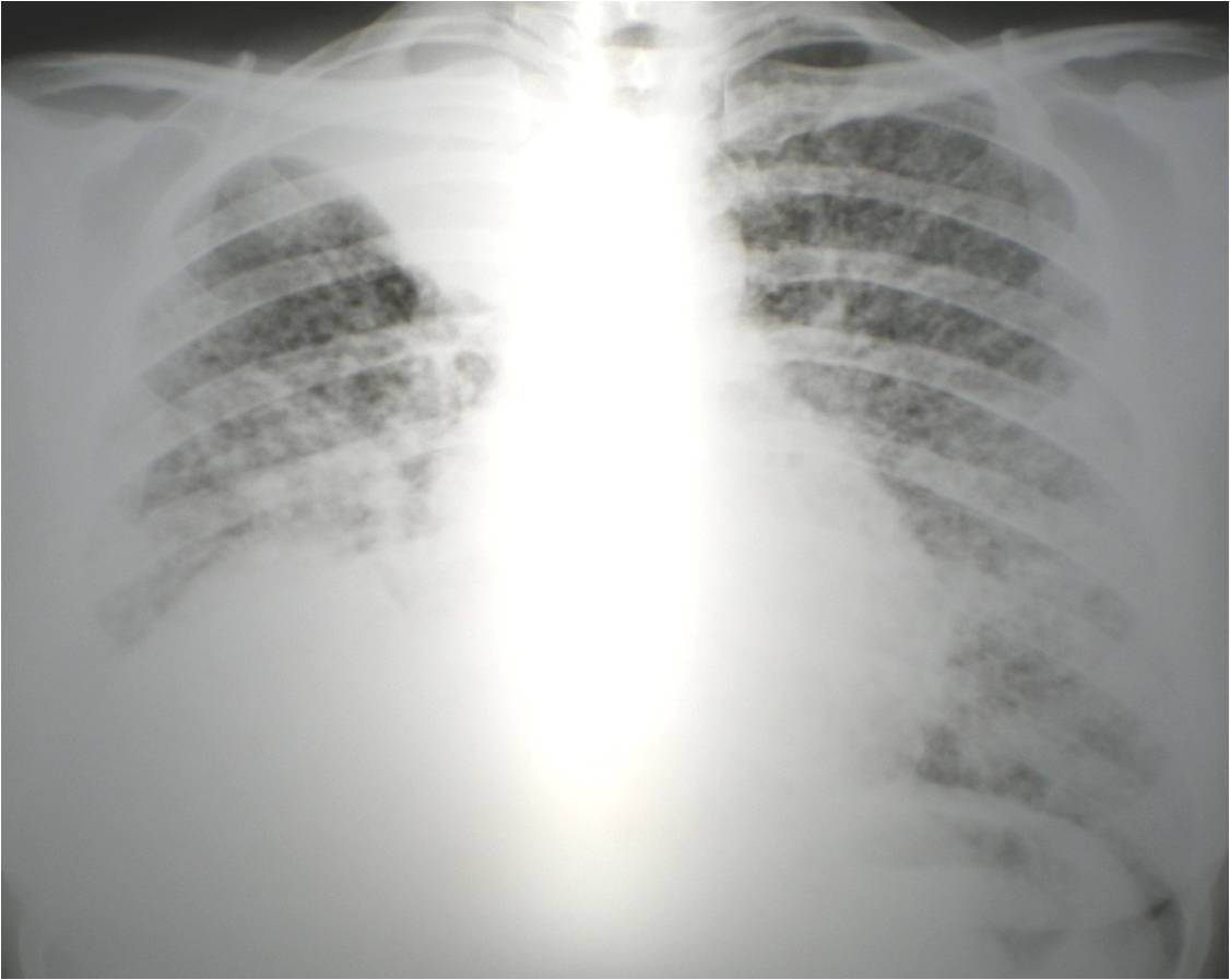
difusos.Insuficiencia respiratoria. Radiografía:
radiopacidadintersticionodulillar difusa, con imagen
radiopaca homogénea en vértice derecho. TC-AR: engrosamiento
nodular de los septos interlobulares e intersticio
peribroncovascular. Se inicia tratamiento para TBC pulmonar.
Evoluciona desfavorablemente con requerimiento de ARM y
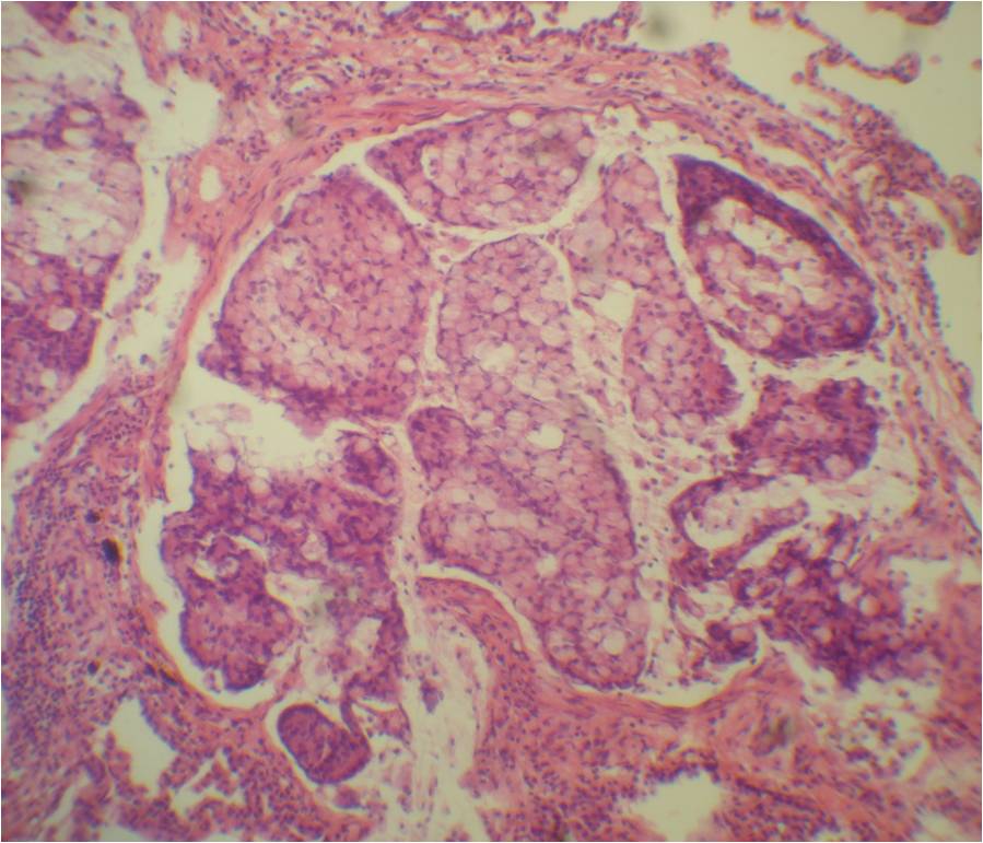
desenlace fatal. En ambos casos se realizó biopsia pulmonar
con diagnóstico de LC. Discusión: Se presentan dos casos de
neumopatía aguda afebril en pacientes jóvenes, cuyas
manifestaciones orientaron al diagnóstico de infección
respiratoria, y evolucionaron rápidamente hacia la
insuficiencia respiratoria refractaria con requerimiento de
ARM y desenlace fatal. Conclusión: La LC pulmonar debe
incluirse comodiagnóstico diferencial ante cuadros de
neumopatía aguda, principalmente cuando la evolución es
desfavorable bajo antibióticos o se han excluido etiologías
más frecuentes.
Palabras claves: linfangitis carcinomatosa pulmonar,
carcinomatosislinfangítica
Abstract
Introduction: Carcinomatous lymphangitis of the lung
accounts for 6-8% of lung´s metastases. There are evidence
that it can be a treatable condition with an impact on
progression of dyspnea and radiographic lesions, and
survival improvement. Two cases are reported, with the aim
of increase the clinical suspicion at compatible cases.
Methods:Case 1: woman 32 years old. Progressive dyspnea and
cough two weeks ago, without antibiotic response. Tachypnea,
increase of respiratory work, basal crackles.Respiratory
failure. Radiography: basal alveolointerstitial opacities.
Treatment for severe community-acquired pneumonia is started.
She evolves unfavorably, with need of MRA and fatal outcome.
Case 2: woman 46 years old. Progressive dyspnea and cough
from one week ago. Tachypnea, diffuse crackles. Respiratory
failure. Radiography: diffuse nodular-interstitialradiopacity,
with radiopacy lesion in right apex. HRCT: nodular
thickening of interlobular septum and peribrochovascular
interstitial. Treatment for tuberculosis of the lung is
started. She evolves unfavorably, with need of MRA and fatal
outcome. In both cases a lung biopsy was performed,
diagnosing carcinomatous lymphangitis. Discussion: Two cases
are reported, with acute afebrilepneumopathyin young
patients, whose manifestations guided to treatment of
respiratory infection, and evolved quickly to refractory
hypoxemic respiratory failure with need of MRA and fatal
outcome.Conclusion: Carcinomatous lymphangitis of the lung
should be included as a differential diagnosis of cases of
acute lung pathology, especially when there was an
unfavorable evolution under antibiotics or have excluded
more common etiologies.
Key words: carcinomatous lymphangitis of the lungs,
lymphangitic carcinomatous.
Imagenes
|